112 a posteroanterior and b lateral chest radiographs of a cardiac resynchronization therapy device crt d system with the pulse generator located in a retromammary position. Scaphoid fractures comprise about 5090 of carpal bone fractures early accurate diagnosis and the introduction of proper treatment results in about 9496 healing rate misdiagnosis may lead to non union avascular necrosis and consecutive osteoarthritis of the wrist joint.

Both Bone Forearm Fracture Radiologypics Com

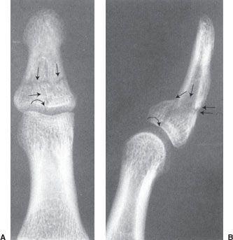
The Radiology Assistant Wrist Fractures

The Monteggiafracture Literature Review And Report Of A New Variant
Note that the right ventricular coil is in a low outflowhigh septal position.

Radiology fracture apex angulation. The knee skyline laurin view is an inferior superior projection of the patellait is one of many different methods to obtain an axial projection of the patella. Le fort fractures are fractures of the midface which collectively involve separation of all or a portion of the midface from the skull basein order to be separated from the skull base the pterygoid plates of the sphenoid bone need to be involved as these connect the midface to the sphenoid bone dorsally. Similar to laws view but cephalocaudal beam makes an angle of 30 degrees instead of 15 degrees stenvers view axio anterior oblique posterior.
The management of these fractures differ greatly from fractures of the other regions and hence are. These fractures are different from tibial shaft fractures as the treatment approach and outcomes are different. The le fort classification system attempts to distinguish according to the plane of injury.
Although thoracic spine pain can be as chronic and disabling as pain in the cervical or lumbar regions of the spine it is less common underreported and less studied in the literature. Sagittal plane of the skull is parallel to the film and x ray beam is projected 15 degrees cephalocaudal schullers or rugnstrom view 30o lateral oblique. Due to its common availability and low costs plain radiography of the wrist.
Subcutaneous edema and effusionhypertrophy of synovial membrane increased density of periarticular soft tissues. Spindle nodular enlengthened morphology with net or confused borders. The preferred view for distinguishing outflow tract and coronary sinus cs positions is the lateral view.
Like tibial plateau fractures fractures of the proximal third of tibia are associated with severe soft tissue injuries. 1 anatomy and imaging of the shoulder joint. The head and the glenoid fossa articulate in the shoulder joint glenohumeral joint.
Laws view 15o lateral oblique. Facing the film and head slightly flexed and. Pradeep chopra in current therapy in pain 2009.
Shaft femur fracture is the fracture of the diaphysis of the femur an area from the subtrochanteric region to supracondylar region. This projection is best suited to patients able to maintain a semi recumbent position on the examination table. The biggest reason for this is that the thoracic region of the spine has less mobility and flexibility than the cervical or lumbar.
Tumefaction of soft tissuesit is assessed with echography or mri conventional x rays arthrography is no longer used yes.
6 Musculoskeletal System Radiology Key

The Radiology Assistant Wrist Fractures
Boxer Fracture Radiology Reference Article Radiopaedia Org